Towards measuring surface dynamics with neutrons
Why?
Surface science has advanced enormously in recent decades, but many scientific questions are not resolved. Examples are spin wave dispersions with experiments contrasting theory or a difference in the potential defining the zero point energy of H adsorbed at Pt and Si surfaces. The surface dynamics of glass formers is under discussion as well as that of self-assembled monolayers. An important aspect in biology is the dynamics of water close to membranes, which strongly depends on the head groups. Finally, understanding the dynamics of lithium and protons in thin films is crucial for the development of batteries, fuel cells, sensors, and biology. Many studies of surface effects rely on molecular dynamics simulations or spectroscopy using charged particles with a limited probing depth (or direct contact with a sample, e.g., atomic force microscopy) possibly biasing the results. Other methods probe macroscopic quantities, like, e.g., viscosity and the use of photons in the visible range is restricted to small momentum transfers and subject to various selection rules. Neutrons interact via the strong force with nuclei and have a spin and thermal energies, which probe the whole Brillouin zone, allowing studies of phonon or magnon dispersions as well as they are directly sensitive to tracer diffusion and vibrational modes.
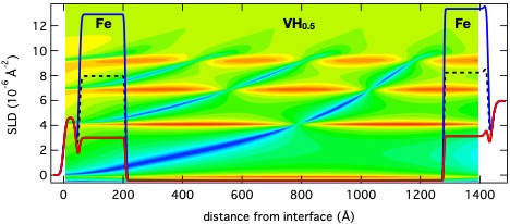
Figure 1. Scattering potential for the resonator. A potential well is formed by vanadium sandwiched between iron layers. The underlying color map depicts the calculated wavefield for different incident beam momenta. The enhancement of the wave function is visible by the red color.
How?
With neutrons in surface science, the density profile across an interface is extracted from specular neutron reflectivity (NR) in a routine way. Only a few studies probe in-plane structures and a very limited number dynamics. The reason for this is the limited brilliance at nowadays neutron sources. One way to overcome this is the use of quantum resonators in order to enhance the signal. We use this method combined with magnetic contrast variation to control the neutron wave amplitude and unambiguously detect incoherent scattering from protons at interstitial lattice sites in a thin film of V, paving the road for future studies of dynamics.
Figure 1 shows the scattering potential (scattering length density (SLD)) for neutrons impinging under grazing incidence onto the sample surface. A layer of vanadium hydride is sandwiched between iron layers and forms a potential well. The film is grown on MgO and protected against oxidation by a palladium and aluminum oxide coatings. For specific incident momenta the neutron wave field (shown by the underlying color map) gets enhanced, similar to light getting guided in an optical fiber, and resulting in an increased scattering probability from inside the resonator.
Tracer diffusion is measured by neutrons via incoherent scattering, which can be related to the Fourier transform of the particle self-correlation function in space and time. Since neutrons scatter from the nucleus, which is much smaller than the wavelength of the neutrons incoherent scattering is isotropic. In order to capture as much as possible of this scattering we have glued the sample onto a well shielded neutron detector, leaving only a small window towards the sample open.
Figure 2 depicts the reflected neutron intensity from the sample plotted together with the signal registered by the detector directly behind the sample. The region of total external reflection is visible for Qz values below about 0.02 Å-1. The dips in the signal are at the positions when the evanescent wave enters the resonator. At exactly these positions the detector catching the incoherent scattering registers neutrons. To proof that the detected signal is indeed originating from the resonator we have magnetized the iron layers and used spin polarized neutrons for the experiment. In this way the walls of the resonator are changed in height for the different spin states of the incoming beam and the resonances shift in position. It is interesting to note that the overall incoherent background can be attributed to the incoherent cross section of magnesium and intensity transmitted through the resonator.
Our measurements demonstrate that the incoherent signal from a thin film of 100 nm can be measured and pave the road towards experiments probing tracer diffusion and dynamics in thin films and at interfaces. Such measurements require high brilliance sources and a dedicated sampler design.
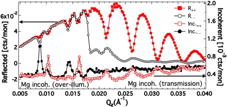
Figure 2. Intensity reflected from the sample surface (left axis) for + and – polarized neutrons plotted together with the signal collected at a detector mounted directly behind the sample to measure the incoherent scattering.
What's next?
As a next step we will include energy analysis into our experiments and measure inelastic and qusielastic scattering to address dynamics in thin films. The proof of principle will be done on a direct geometry chopper time of flight spectrometer with flexible incident wavelength resolution, a large detector area (on the order of (π/2) and comparable to our previous experiment), and a monochromatic beam. With the choppers open, the incoherent signal can be captured and the sample aligned. Once aligned, the beam might be chopped and measurements will be performed on the energy gain side to maximize the pulse repetition rate. In the further future we expect considerable gain factors if a dedicated beam line optimized in all respects would become available.
Who?
This work was conducted by a team of researchers from Uppsala Univerisity at the national infrastructure Super ADAM at the Institute Laue-Langevin (ILL) in Grenoble (France) in collaboration with scientists from the NIST center for neutron research (NCNR), Gaithersburg, MD (USA) and the Petersburg Nuclear Physics Institute, Gatchina (Russia).
Contact:
Max Wolff, Uppsala University
max.wolff@physics.uu.se
Further information: PHYSICAL REVIEW LETTERS 123, 016101 (2019)
This project is funded by the Swedish Research Council. The experimental data were collected on the Super ADAM beamline at the Institut Laue-Langevin (ILL), Grenoble, France.